
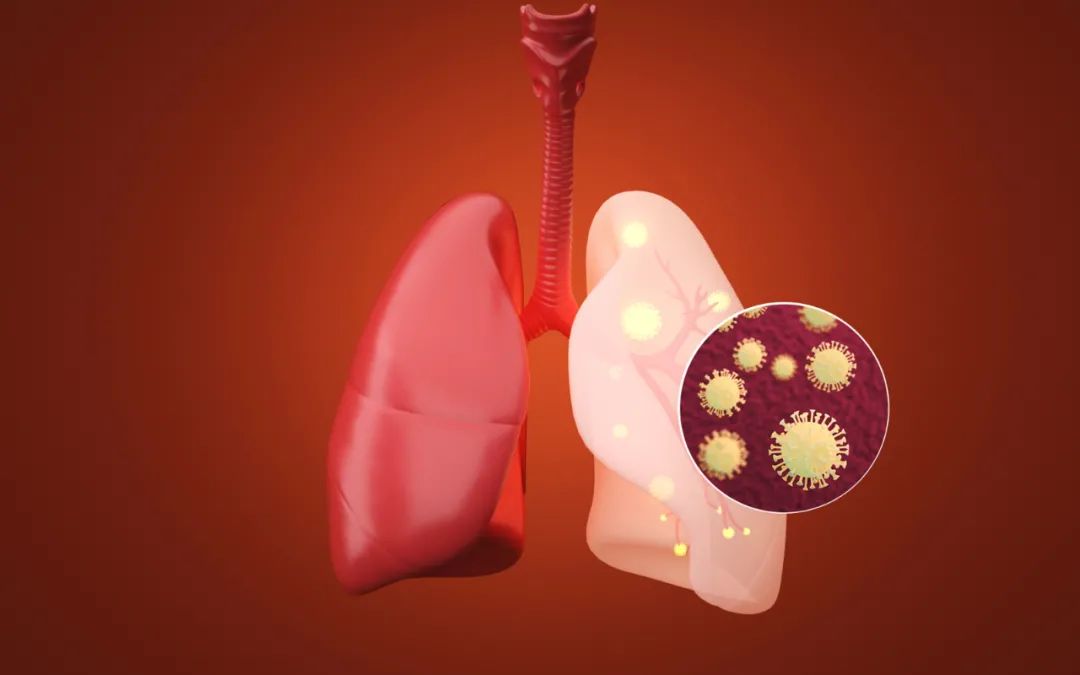
2018-07-02
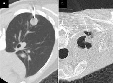
肺结节的CT影像学分类(根据结节的密度/实性比例分类)
● 实性结节(Solid nodule )
1.按大小分类:≤4mm,>4-6mm,>6-8mm,>8mm
2.按病变个数:多发,单发

● 亚(部分)实性结节(sub-solid, or part solid nodule)
1.按实性成分大小:≤5mm,>5mm
2.按病变个数:多发,单发

● 纯毛玻璃样结节( Ground glass opacity,GGO )
1.按GGO大小:≤5mm,>5mm
2.按病变个数:多发纯GGO(Multiple GGO),单发纯GGO

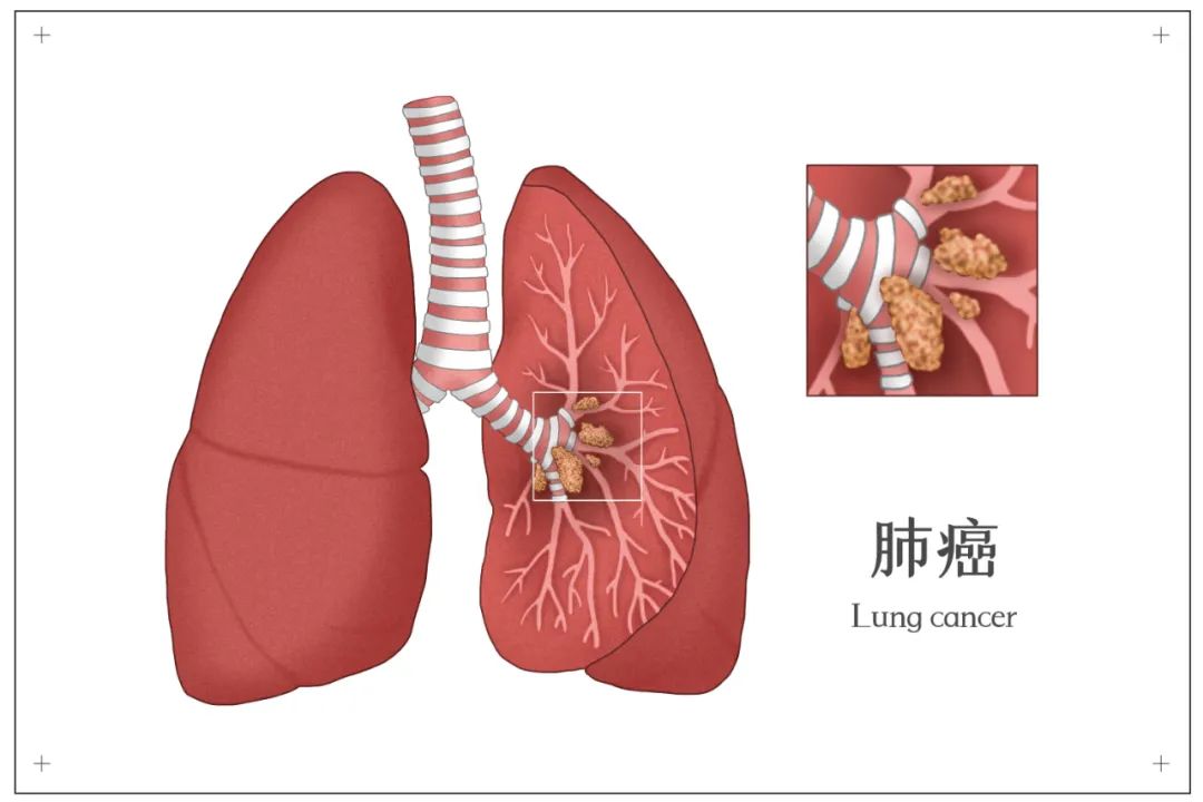
# 非实性结节(Nonsolid nodule,NS)包括亚实性和毛玻璃样结节
作者:杨学宁
【参考文献】
The IASLC Lung Cancer Staging Project Proposals for the Revisions of the T Descriptors in the Forthcoming Eighth Edition of the TNM Classification for Lung Cancer


推荐阅读
文章评论
注册或登后即可发表评论
登录/注册
全部评论(0)