
2019-04-13
作者:广东省肺癌研究所 杨学宁 & LAMP
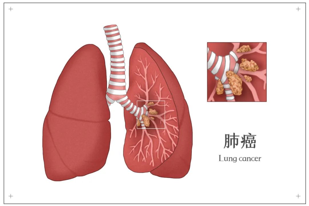
内镜超声下淋巴结采样应用于肺癌的纵隔再分期
245. Endosonography With Lymph Nodes Sampling for Restaging the Mediastinum in Lung Cancer: A Systematic Review and Meta-Analysis
Jiang Long1, Wenhua Liang1, Kassem Harris2, Lonny Yarmus3, Weizhe Huang1, *Jianxing He1
1First Affiliated Hospital of Guangzhou Medical University; State Key Laboratory of Respiratory Disease, National Clinical Research Center for Respiratory Disease, Guangzhou Institute of Respiratory Disease, Guangzhou, China, Guangzhou, China; 2Westchester Medical Center, New York Medical College, New York, NY;3The Johns Hopkins University, Baltimore, MD
Objective: Mediastinal restaging after induction treatment is still a difficult and controversial issue. We aimed to investigate the diagnostic accuracy of endobronchial ultrasound-guided transbronchial needle aspiration (EBUS-TBNA) and endoscopic ultrasound-guided fine needle aspiration (EUS-FNA) for restaging the mediastinum after induction treatment in patients with lung cancer.
Methods: Embase and PubMed databases were searched from conception to July 2018. Data from relevant studies were analyzed to assess sensitivity and specificity of EBUS-TBNA and EUS-FNA, and to fit the Hierarchical Summary Receiver-Operating Characteristic (HSROC) curves.
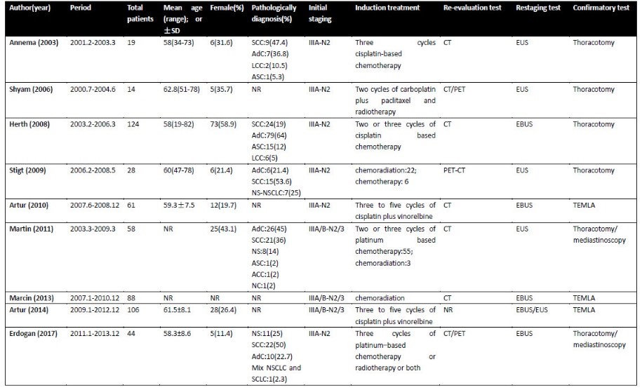
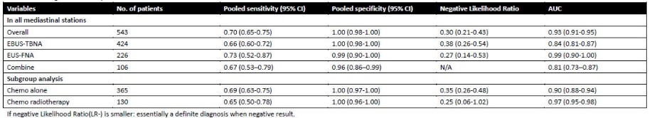
Results: A total of nine studies consisting of 542 patients fulfilled the inclusion criteria. All patients were restaged by EBUS-TBNA, EUS-FNA or both. Negative results were confirmed by subsequent surgical approaches. There were no complications reported during any endosonography approaches reviewed. The pooled sensitivities of EBUS-TBNA and EUS-FNA were 66%(95% CI, 60%-72%) and 73%(95% CI, 52%-87%), respectively; and specificities were 100%(95% CI, 98%-100%) and 99%(95% CI, 90%-100%), respectively. The area under the HSROC curves(AUC) were 0.84(95% CI, 0.81-0.87) for EBUS-TBNA and 0.99(95% CI, 0.98-1) for EUS-FNA. Moreover, for patients who received chemotherapy alone, the pooled sensitivity of endosonography with lymph node sampling for restaging was 69% (95% CI, 63%-75%), and specificity was 100% (95% CI, 97%-100%); and for patients who received chemoradiotherapy, the results seemed similar with sensitivity of 65% (95% CI, 50%-78%) and specificity of 100% (95% CI, 96%-100%).
Conclusions: Endosonography with lymph node sampling is an accurate and safe technique for mediastinal restaging of lung cancer. For nondiagnostic results, a further more invasive approach should be thoroughly considered.


推荐阅读
文章评论
注册或登后即可发表评论
登录/注册
全部评论(0)